TMJD Research
- Mark Stuart Senzig
- 6 days ago
- 6 min read
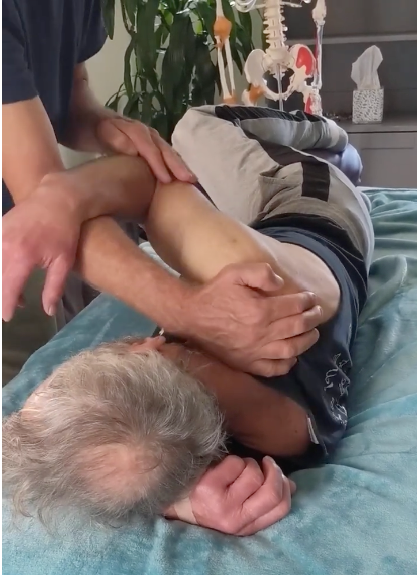
Current scientific research increasingly views temporomandibular disorder (TMD) as a condition involving the jaw joint, masticatory muscles, cervical spine, nervous system, breathing mechanics, and motor-control strategies rather than simply a local jaw problem. Researchers have found that many individuals with TMD demonstrate altered jaw movement, reduced mandibular opening, impaired coordination between opening and closing muscles, increased pain sensitivity, and measurable dysfunction in the cervical region. This broader understanding is important because clinicians frequently encounter patients whose jaw symptoms persist despite local treatment directed only at the temporomandibular joint (TMJ). Modern evidence suggests that restrictions affecting mandibular motion often originate from a combination of joint mechanics, muscle imbalance, cervical dysfunction, postural adaptations, and central nervous system influences.
Research by César Fernández-de-las-Peñas has contributed substantially to understanding the relationship between TMD and cervical dysfunction. His work has demonstrated that patients with chronic TMD frequently present with reduced cervical range of motion, increased tenderness in cervical muscles, altered pressure pain thresholds, and impaired neuromuscular control. These findings support the concept that the jaw and cervical spine function as an integrated biomechanical and neurological system. The trigeminal nerve and upper cervical nerves converge within the trigeminocervical nucleus of the brainstem, creating a mechanism through which dysfunction in the neck may influence jaw pain and vice versa.
Recent studies continue to show that cervical mobility limitations are common in patients with TMD. Researchers including Deborah Armijo-Olivo have reported associations between TMD and reduced upper cervical mobility, particularly involving the atlanto-occipital and atlantoaxial joints. These upper cervical segments contribute significantly to head positioning and mandibular mechanics. When upper cervical extension becomes restricted, patients often compensate through lower cervical extension and forward-head posture. This compensation can alter resting mandibular position and increase loading of the temporomandibular joint and associated musculature.
Muscle imbalance remains one of the most consistent findings in TMD research. The masseter and temporalis muscles are frequently overactive in symptomatic individuals. Electromyographic studies have shown increased resting activity and altered recruitment patterns within these muscles compared to healthy controls. Researchers such as Paulo César Rodrigues Conti have demonstrated that excessive masseter activity may contribute to increased joint loading, pain during chewing, and reduced mandibular excursion. When these muscles remain chronically active, they may restrict jaw opening and create a sensation of stiffness or locking despite the absence of structural joint pathology.
The temporalis muscle deserves particular attention because of its dual role in jaw function and cranial stability. Excessive temporalis activity is commonly observed in individuals who clench their teeth, experience emotional stress, or demonstrate altered occlusal habits. Research suggests that chronic temporalis hyperactivity can influence mandibular resting position and contribute to reduced opening range. Furthermore, temporalis tenderness is frequently associated with tension-type headaches, creating a complex overlap between headache disorders and TMD symptoms.
The lateral pterygoid muscle continues to attract significant research interest. Historically considered difficult to evaluate clinically, advances in imaging and electromyographic investigation have improved understanding of its role. Researchers including Jeffrey P. Okeson have emphasized the importance of the lateral pterygoid in disc-condyle coordination. Dysfunction or excessive activity within this muscle may contribute to abnormal disc movement, joint clicking, and impaired mandibular translation. Rather than functioning solely as a jaw-opening muscle, the lateral pterygoid participates in complex coordination of mandibular motion. Altered recruitment may lead to inefficient movement patterns and restricted range of motion.
The medial pterygoid often receives less attention but plays a substantial role in TMD-related movement restriction. Working synergistically with the masseter, it contributes to mandibular elevation and stabilization. Increased tone within the medial pterygoid may limit contralateral excursion and contribute to restricted opening. Recent imaging studies suggest that muscular adaptations within the medial pterygoid may accompany chronic pain conditions, creating persistent movement limitations even after symptoms improve.
Research by Frank Lobbezoo has advanced understanding of bruxism and its relationship to muscular overload. Bruxism is no longer viewed solely as a dental issue but rather as a multifactorial behavior involving sleep physiology, stress responses, and central nervous system regulation. Chronic clenching and grinding increase loading of the masseter, temporalis, and pterygoid muscles, which may contribute to restricted jaw motion and altered muscle recruitment patterns. Importantly, researchers now recognize that not all TMD patients exhibit bruxism and not all bruxers develop TMD, highlighting the complexity of these relationships.
The suprahyoid and infrahyoid muscle systems are increasingly recognized as important contributors to jaw dysfunction. These muscles coordinate mandibular depression, swallowing, tongue movement, and cervical stability. Research by investigators such as Marina von Piekartz has emphasized the role of the hyoid apparatus in cranio-cervical function. Altered hyoid positioning may influence mandibular mechanics, tongue posture, airway function, and cervical muscle recruitment. Dysfunction within these systems may contribute to restricted jaw opening and impaired movement coordination.
Forward-head posture remains one of the most studied cervical influences on TMD. Researchers including Gwen Jull have demonstrated that chronic forward-head posture is associated with reduced deep cervical flexor function and increased activity of superficial cervical muscles. As the head migrates forward, the mandible often assumes a posteriorly displaced resting position. This alters loading within the TMJ and increases activity of the suboccipitals, sternocleidomastoid, upper trapezius, and masticatory muscles. Over time, these adaptations may reduce mandibular mobility and contribute to persistent symptoms.
Deep cervical flexor dysfunction has become a particularly important research topic. Studies have consistently shown reduced endurance and impaired activation of longus colli and longus capitis in individuals with TMD. When these stabilizing muscles underperform, larger superficial muscles such as the sternocleidomastoid become dominant. This altered motor-control strategy changes head posture and may indirectly influence jaw mechanics. Modern rehabilitation programs increasingly incorporate deep cervical flexor training alongside jaw exercises to address these interconnected dysfunctions.
The sternocleidomastoid muscle has emerged as a major player in the cervical-TMJ relationship. Multiple studies have demonstrated increased tenderness, trigger point prevalence, and altered activation patterns within this muscle among individuals with TMD. Because the sternocleidomastoid influences head position and proprioception, excessive activity may alter mandibular mechanics and contribute to symptoms during chewing, speaking, or prolonged postural tasks.
The upper trapezius and levator scapulae also contribute to movement restriction. Research examining cervical muscle activity in TMD populations frequently identifies increased resting tone and reduced relaxation capacity in these muscles. Elevated shoulder girdle tension may alter cervical mechanics and contribute to compensatory movement strategies affecting mandibular motion. Rather than viewing jaw dysfunction in isolation, contemporary research supports evaluating the entire cranio-cervical-shoulder complex.
Researchers such as Peter Svensson have expanded understanding of pain mechanisms in TMD. His work highlights the role of central sensitization, whereby the nervous system becomes increasingly responsive to sensory input. In such cases, movement restriction may not be entirely mechanical. Patients may demonstrate protective muscle guarding, altered motor control, and reduced movement variability despite minimal structural pathology. This finding helps explain why some individuals continue to experience restricted jaw motion even after local tissue healing has occurred.
Breathing mechanics have become another emerging area of interest. Investigators studying airway function and craniofacial development have reported associations between mouth breathing, altered tongue posture, forward-head posture, and TMD symptoms. Chronic mouth breathing may encourage cervical extension, elevated accessory breathing muscle activity, and altered mandibular positioning. These adaptations can increase tension throughout the suprahyoid, infrahyoid, scalene, and sternocleidomastoid systems, potentially limiting efficient jaw movement.
Research involving movement analysis has shown that healthy mandibular motion requires precise coordination between the temporomandibular joint, cervical spine, tongue, hyoid apparatus, and masticatory muscles. Studies using three-dimensional motion capture demonstrate that jaw opening is accompanied by subtle cervical extension while jaw closing is associated with cervical flexion. When cervical mobility is restricted, these normal coupling patterns may become disrupted, resulting in compensatory movement strategies and reduced efficiency.
Current evidence therefore supports a regional interdependence model. According to this framework, TMJ dysfunction is influenced not only by local joint structures but also by cervical mobility, neuromuscular control, postural adaptation, breathing mechanics, and central pain processing. Researchers including Fernández-de-las-Peñas, Armijo-Olivo, Jull, Lobbezoo, Svensson, Conti, Okeson, and von Piekartz have collectively demonstrated that successful management of TMD often requires attention to both jaw-specific impairments and dysfunctions arising from the cervical spine and surrounding musculature.
For manual therapists, the most important modern conclusion is that restricted mandibular opening, deviation, lateral excursion limitations, and painful chewing are rarely caused by a single tight muscle or isolated TMJ problem. Instead, movement restrictions frequently reflect interactions among the masseter, temporalis, pterygoids, suprahyoids, deep cervical flexors, sternocleidomastoid, upper trapezius, levator scapulae, suboccipitals, tongue musculature, breathing patterns, cervical joint mobility, and nervous system sensitivity. The strongest trend in current research is moving away from a purely local jaw model toward a comprehensive cranio-cervical movement system model in which the neck and jaw function as inseparable partners in normal movement and dysfunction.


Comments